A chiropractic case review
Cervical Spondylotic
Myelopathy:
A Review and Case Report.
Jeffrey R. Cates, DC, DABCO*
Morris Marc Soriano, MD†
*Private practice of chiropractic
E-mail
Dr. Cates - cates@essex1.com
![]() Click logo
to go to Cates homepage.
Click logo
to go to Cates homepage.
200 North 6th Street
Oregon, IL 61061
(815) 732-2686
†Private practice of neurosurgery
1021 N. Mulford Rd.
Rockford, IL 61108
(815) 395-1991
Abstract - JMPT Vol 18, Number 7 September 1995; Pgs 471-475: SSN 0161-4754
Objective: To present the clinical and neurological features of cervical spondylotic myelopathy by review and case presentation.
Clinical Features: Cervical spondylotic myelopathy (CSM) is a condition where the vascular and neural structures are compressed by bony spurring and soft tissue hypertrophy which causes ischemic damage to the spinal cord. While cervical spondylotic myelopathy is the most common cord disorder in older adults, the diagnosis is often missed since the initial symptoms are subtle and the condition usually presents with associated conditions such as nerve root involvement.
Intervention: The patient was referred to a neurosurgeon for a posterior decompressive laminectomy. The advancing symptoms of CSM were apparently halted by the surgery in this case until complication from a fall resulted in quadriplegia.
Conclusion: Appropriate testing can aid differential diagnosis of the condition and expedite appropriate management of the condition. Treatment may include surgical cervical decompression of the involved area. An untreated progressive spondylotic myelopathy may cause permanent neurologic damage to the spinal cord. It is the authors intent to call attention to the clinical signs and treatment of this underdiagnosed condition.1
Key Indexing Terms: Spondylotic Myelopathy, Cervical, spondylosis, Spinal Stenosis, Myelopathy, Spondylitis.
Introduction:
Stumpell presented the first case of cervical spondylotic myelopathy in 1888 2, however, detailed research into the mechanism of injury was documented by Brain, Northfield and Wilkinson in 1952.2, 3 Cervical spondylotic myelopathy (CSM) is common in older adults with degenerative spondylosis of the cervical spine.1 There is a good chance that the practicing chiropractor may encounter several of these cases each year. Since the early symptoms of CSM are subtle, the diagnosis may be missed. A case study and review is presented to inform the doctor of the condition and aid in diagnosis and appropriate treatment.
Case Study:
CHIROPRACTIC: The patient, a 55-year-old white illiterate male, presented himself to the chiropractic office in mid-February 1994 complaining of hand weakness, neck, shoulder and right arm pain. The patient had a long history of alcohol and tobacco use. Prior history included bilateral ulnar nerve release in July 1990. Cervical disc disease with polyneuropathy and radiculopathy was also diagnosed in 1990. An MRI at that time revealed spondylotic changes from C5 to C7, however, the cord was reportedly normal. He reportedly tried a course of hospital rehabilitation in 1991 with no noticeable improvement. The condition was aggravated a week prior to presentation. He reported that the arm and hand became markedly weaker and more painful on awakening one morning, and that the condition was aggravated by lying on his back. The patient claimed it felt better when lifting his hand and arm over his head.
On examination, it was most evident that the patient had difficulty holding a pen. There was notable wasting of the intrinsic hand muscles with diffuse weakness of bilateral upper extremity musculature. Bilateral upper extremity reflexes were depressed. Hyperreflexia was noted at the knee 3+/3+; ankle reflexes were 2+/2+. Babinski's test revealed great toe reflexes that were equivocal / upgoing bilaterally. There was a notable asymmetrical loss of cool sensation and vibratory sensation in the lower extremities most notably on the right side. This was thought to be significant as a gradient symmetrical loss of sensation would tend to indicate a peripheral polyneuropathy or benign senile degeneration while the asymmetric loss of sensation tends to indicate a more selective lesion such as CSM. His gait was normal on initial chiropractic examination. X-ray examination showed marked spondylotic stenosis of C5 to C7 with the narrowest spondylotic AP diameter of only 10 mm and a body / canal ratio of 0.59 at C6. (See figure 3).
Fig. 3 - The patients lateral x-ray depicted marked spodylosis at the C5 - C6 level. With only 10 mm of space available to the spinal cord and a 0.59 Pavlov ratio, cord compression is likely.
No chiropractic care was rendered in this instance, and the patient was immediately referred to a neurosurgeon for further evaluation for suspected cervical spondylotic myelopathy.
SURGICAL: After extensive neurologic, x-ray and hematological evaluation, it was confirmed that this gentleman was suffering from a compressive cervical myelopathy secondary to degenerative spurs in the cervical spine from C3 to C6. During the work-up period, additional symptoms related to the CSM presented, such as urinary dysfunction and an abnormal gait. The patient was taken to surgery in May of 1994 for a posterior decompressive laminectomy. A skin incision was made over the spinous processes down to the ligamentum nuchae and the muscles stripped free from the spinous processes and laminae bilaterally from C4 to C6. Prominent portions of the laminal and spinous processes of the same vertebra were removed. On examination, it was found to be extremely tight at the C4-C5 junction with tremendous cord pressure and no significant vascular pulsation of the cord noted until after the decompression, at which time, significant pulsations of the cord were noted. Care was taken to leave the laminae so as to prevent any swan-neck deformity. The remaining portion of the procedure was performed in an uneventful fashion. The patient did well from his surgical intervention. After surgery, the patient did note improvement in his ambulation and stability of his gait. He also noted improvement of his urinary function and subjectively experienced increased strength in his lower extremities.
OUTCOME: Four months following the surgery and discharge form the hospital, the patient had stabilized with no progression of the disease. Unfortunately, the patient fell at home striking his head quite severely on the floor in September of 1994. The patient suffered acute quadriplegia at the C4 level and was admitted to the hospital. Review of the x-rays showed no hematoma or fractures in the spinal canal. There was evidence of intrinsic cord damage on the MRI scan at the C3-4 level from a large posterior spur that had been impaled into the cord.
In this patient's case, it appears he had done extremely well following his laminectomy and was in fact experiencing improvement. Unfortunately, the posterior laminectomy does have its limitations in that it does not attempt to relieve the pressure from the anterior spurs impinging upon the anterior portion of the dural sac. Anterior decompression was considered in this gentleman, but, because of the multiple levels and debilitated condition, it was felt that a simpler, less stressful posterior decompression would be the operation of choice. Currently the patient is in rehab with a feeding tube and tracheostomy in place.
Discussion:
Current literature indicates that although spondylotic myelopathy is the most common type of cord disorder in the elderly, it is probable that a good many of these cases go undiagnosed until later stages of the disease 4, 5
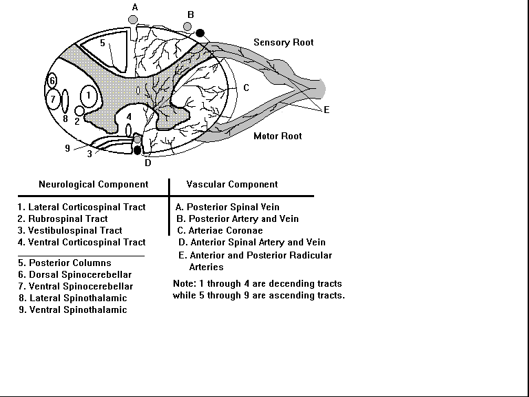
Cause: In CSM the spinal canal becomes stenotic and the spinal cord and vascular structures are pinched and irritated by bony spur formation, thickened ligaments, dural root adhesions, venous congestion and dynamic posture changes.6, 7 Disc degeneration with spondylotic reactive change is the major factor in development of CSM.6 Chronic pressure on the cord and vascular structures causes slow but progressive demyelination and / or ischemia with associated symptoms that vary according to the location and degree of cord damage. Current theory would indicate ischemic compression of the anterior spinal artery, paired posterior spinal arteries and radicular arteries which lie in the neural foramen of the cervical vertebrae as the primary cause of the myelopathy.4, 7, 8 Ischemic damage is often greatest in the anterolateral white and the central gray matter of the cord,5 with a substantial degree of damage occurring when the neck is held in unusual posture while sleeping. The C5 - C6 region of the cord is most vulnerable to vascular ischemia.6 The anterior artery supplies 60% - 70% of the cord, mainly the anterior horns and the pyramidal and spinothalamic tracts, while the posterior spinal arteries supply the posterior horn and dorsal columns.3, 6 The pyramidal tracts are often the first to be involved, and often most severely, next the spinothalamic tracts and later possibly the posterior columns.9 Vascular embarrassment is thought to occur to the small radicular arteries and vessels on the cord surface.6 Acute injuries, such as a whiplash or a fall, may initiate or exacerbate symptoms in a patient with cervical spondylosis.6, 8
The types of CSM are classified according to the anatomical region involved.3, 6, 7, 8
1. Lateral - Involves damage to the radicular component involving the nerve root. Pure lateral type CSM exhibits no long tract damage.
2. Medial - Involves damage to the spinal component. Typically causes asymmetric long tract signs and symptoms that vary according to the specific tracts damaged.
3. Combined - Involves varying degrees of both the lateral and medial types which includes both nerve root and spinal cord damage. This is by far the most common.
4. Vascular - Involves a rapid onset with-out pain which develops after little-to-no trauma. This form is consider the least common.
Clinical Picture: The presenting symptoms will vary depending on the site of ischemic damage to the cord. Therefore, a working knowledge of the vascular and neurological anatomy of the cervical spine is needed to evaluate the disorder. (See figure 1)
Fig. 1 - Tracts & neural structures depicted on left, vascular structures on right.
 The average onset is reported to be in
the mid 50's 10 , with a higher incidence reported in
males. A common complaint is mild neck discomfort. Often the
patient will report no pain, only "clumsy or weak"
hands or feet that may be hypersensitive. 5 Tingling
fingers and leg stiffness are common complaints.11 Lhermitte's sign, a shocking sensation
radiating out the upper or lower extremity with neck flexion, may
be reported in some cases. With vestibulospinal tract
involvement, a gait disturbance may be noted. The patient may
walk bent forward with a wide-based gait due to loss of
coordination. Examination may reveal subtle loss of posterior
column function; therefore deep touch, vibration, joint
proprioception and two-point sensations may be disturbed.7 With damage to the spinothalamic tract,
the patient may claim they can't feel a cold floor or hot water
in the tub. With damage to the corticospinal tracts, spasticity
with motor weakness of the upper or lower extremities may be
noted; however, motor signs are often asymmetric.2 Atrophy of the intrinsic hand musculature
may present. Combined CSM is by far the most common type of
damage seen. Clinically patients with combined CSM may present
with what appears to be simple cervical nerve root involvement
and often are misdiagnosed as such. It is interesting to note
that the motor and sensory component of combined CSM may
deteriorate or improve at different rates. 2 There
is usually relative sparring of the anterior tracts.7 The clinical outcome may vary from minor
neurologic dysfunction occurring over a long period of time to
catastrophic acute deterioration presenting within a very short
time period.4 (See table 1.)
TRACT FUNCTION
| 1. Lateral Corticospinal | -Voluntary motion. |
| 2. Rubrospinal | -Muscle tone and synergy. |
| 3. Vestibulospinal | -Balance reflex. |
| 4. Ventral Corticospinal | -Voluntary motion. |
| 5. Posterior Columns | -Vibration, pasive motion, joint & two point discrimination. |
| 6. Dorsal Spinocerebellar | -Reflex proprioception. |
| 7. Ventral spinocerebellar | -Reflex proprioception. |
| 8. Lateral Spinothalamic | -Pain and temperature sensation. |
| 9. Ventral Spinothalamic | -Light touch sensation. |
Table 1
Spinal cord tracts and associated neurologic functions are listed in table 1. The number sequence corresponds to the numbered neurologic structures depicted in figure 1.
Testing: Check two-point sensation, vibratory sensation, temperature discrimination and joint proprioception. Testing of fine-joint proprioception can be achieved by asking the patient to close their eyes and indicate if you are moving their middle toe up or down. Advanced disease to the corticospinal tract can cause a positive Babinski's or Hoffman's sign. Lesions above C6 may cause an inverted radial jerk reflex on percussion over the radial tubercle.12 Clonus may be noted on dorsiflexion of the foot or flexion of the wrist. Fasciculations of the muscles may be present and can be seen, felt and reportedly heard by auscultated of the involved muscle.10 The patient may display signs of spasticity, increased muscle tonus and hyperreflexia. 2, 12, 13
Lower motor neuron signs at the level of the damage with upper motor neuron signs below the level of injury are commonly noted. There may be a gradient loss of temperature and vibratory sensation in the extremities associated with damage to the anterior portion of the posterior columns.2
A mild gradient loss of vibratory sensation and proprioception can be normal as some degree of senile posterior column degeneration is expected with aging. Most often, the ischemic damage causes mixed root and cord symptoms, so it is very easy to mistake these lesions for simple cervical nerve root damage only, especially when the early cord symptoms are subtle.
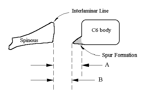
X-ray findings: Cervical x-rays often show reduced space available to the spinal cord; this may be congenital or acquired from progressive spondylosis. 14 Measure the space available to the cord using one of the following methods: the developmental AP (anteroposterior) diameter, the spondylotic AP diameter or Pavlov's ratio.
The developmental AP diameter, or "pre-existing" AP diameter, has been described as the diameter of the canal as measured from dorsal aspect of the mid-vertebral body to the interlaminar line. The average normal C4 - C6 AP diameter was reported by Wilkinson et al. to be 18.5 mm. Spinal stenosis is considered with less than 13 - 14 mm. This measurement is taken on lateral cervical films with a 72" focal-film-distance.15
The Spondylotic AP or "absolute" AP diameter is made by measuring from the posterior spur to the interlaminar line. If the smallest spondylotic AP diameter is 13 mm or greater in the adult, it is unlikely that the spondylotic changes are the cause of the cord compression. This measurement is also taken on lateral cervical films with a 72" focal-film-distance. 6, 12, 15, 16.
Pavlov's Ratio is made by measuring the distance from the middle of the posterior aspect of the vertebral body to the laminar line and comparing it to the mid-vertebral body measurement. A ratio less than 0.82 is indicative of stenosis with 92% accuracy. This technique has the advantage of being accurate independent of the focal-film-distance as the ratio system is unaffected by magnification and is reportedly more than 2 1/2 times more sensitive than the conventional methods listed above. 16
When evaluating any imaging study, including x-rays, one must take into consideration that the neck is a mobile system, and various neck movements change the momentary space available to the cord. For example, due to the folding of ligamentous structures and vertebral positional changes, the spinal dural canal is 2 - 3 mm smaller in extension while the spinal cord thickens in diameter and the intervertebral foramen are smaller by 20 - 30%. 3, 9, 17
Fig. 2
Two methods of assessing space available to the spinal cord using lateral cervical x-rays are shown in figure 2. Note that measurements for method 1 are valid when using a focal film distance of 72 inches.

Method 1
A-Normal C6 D.A.D. Developmental AP diameter is 18 mm.
B-At risk -Stenotic C6 S.A.D. (Spondylitic or "Absolute" AP diameter) is less than 12 - 13 mm.
-An S.A.D of less than 10 mm indicates cord compression.
Method 2
Pavlov's Ratio. The AP diameter of the vertebral canal is divided by the AP diameter of the body. A ratio of < 0.82 indicates stenosis.
Special Test: Additional imaging such as CT with metrizamide myelography or MRI may be utilized to further evaluate the degree of cord compression. 1, 6, 12 Bony transverse bars are found in 80% of case of CSM.9 High resolution spinal angiography may help define levels of greatest vascular compromise.6 Electromyographic studies (EMG testing) can help sort out cord-vs-root damage by H reflex and reflex pattern evaluation. Testing somatosensory evoked potentials (SSEP's) can help evaluate posterior column function. SSEP's are commonly altered with CSM. 2, 7
Laboratory Findings: Neuro-sensitive lab tests can help differentially diagnose the various types of neuropathy. A sensory motor neuropathy blood profile may include testing of SGPT (alanine aminotransferase). MAG (myelin associated glycoprotein) may be elevated in cases of chronic slowly progressive demylinating diseases such as CSM. GM 1 (ganglioside monosialic acid) Anti GM 1 anti bodies are present in patients with sensori- motor, motor neuropathies and lower motor diseases. High anti-sulfatide titers are associated with sensory axonal polyneuropathies. High HU (anti-neuronal nuclear antibody) titers are often associated with sensory neuropathies secondary to small cell lung carcinoma. Increased protein in the cerebrospinal fluid has been associated with CSM.5 As with all lab tests, clinical correlation is important.
Differential Diagnosis and Complications: The differential diagnosis must take into account the possibility of both the myelopathy and radiculopathy. Differential diagnosis for CSM includes: amyotrophic lateral sclerosis, multiple sclerosis, cord tumors, arterial-venous malformations, Vitamin B12 deficiency, metastatic cord compression, atlantoaxial subluxation, thoracic outlet syndromes, cervical disc lesions, Charcot-Marie-Tooth disease, carpal and cubital tunnel syndrome.8 Some cases of spondylotic myelopathy may be compounded by alcohol, diabetes, mononeuritis multiplex or peripheral nerve entrapment syndromes 12 creating complexed polyneuropathies that are difficult to diagnosis and treat.
Treatment: Patients with symptoms consistent with cervical myelopathy should be evaluated by a neurologist. Conservative measures may include heat, massage and / or cervical traction with immobilization in a cervical collar for several months 5 ; however, only 50% of patients improve initially with conservative treatment 7 . The patient may consider sleeping with a soft collar to avoid sleeping postures that may aggravate the condition. While mobilization and manipulation procedures are useful in cases of neck and arm pain due to disc and soft tissue injury or
dysfunction,18 these procedures are controversial when CSM is involved. Most authors feel manipulation is inadvisable while others believe it may be indicated in select cases, usually distal to the region of the CSM. 3, 4, 8 Surgical decompression is indicated in cases of confirmed cord compression with progressive symptoms and usually has a better out-come than conservative approaches 1, 3. Either an anterior discectomy with removal of bony bar formation or a posterior approach involving a laminecotomy or laminoplasty of the stenotic region, and in some cases section of dentate ligaments, are commonly performed.19 The surgical approach is often dictated by the number of vertebral levels involved and the site of damage.13, 20, 21, 22, 23 The prognosis is poorer with advancing age, rapidly progressive degeneration, sphincter dysfunction, posterior column dysfunction and lower extremity weakness 3, 5. No treatment methods have been effective in reversing neurological damage existing more than one year 3, even surgery offers only the hope of halting the progression of the disorder. Early diagnosis of CSM is important since failure to recognize and properly treat the disorder can result in permanent cord damage with irreversible related neurologic deficits.13
Conclusions: CSM is the most common cord disorder in the elderly. Many cases are misdiagnosed or go undiagnosed due to its subtle and varying presentation. By observing and testing a patient in an office setting, a doctor can establish an early differential diagnosis for spondylotic myelopathy. When spondylotic myelopathy is suspected, the doctor should seek early confirmation by referral to a neurologist. Trauma prior to or following surgery can create substantial complications. Early identification, confirmation and management may prevent progressive irreversible neurologic deficits.
E-mail
Dr. Cates - cates@essex1.com ![]() Click logo
to go to Cates homepage.
Click logo
to go to Cates homepage.
Acknowledgment page
The authors would like to thank Robert Porter, MD and the Swedish American Hospital E.R. staff for helping with the literature search, and Christina B. Jensen, DC for her assistance with photography and proof reading. Thanks to Dale Hoppe for his assistance in the final editing. The authors also express appreciation to Specialty Laboratories and Genica Corporation for their contributions of laboratory information on sensory motor neuropathy blood profile data.
References
1. Long, DM. Lumbar and cervical spondylosis and spondylitic myelopathy. Current Opinions in Neurol Neurosurgery 1993; (Aug) 6 (4): 576-80.
2. Tang XF, Ren ZY. Magnetic transcranial motor and somaosensory evoked potentials in cervical spondylitic myelopathy. Chinese Medical Journal 1991; (May) 104 (5): 409-15.
3. Skogsberg, DR. Chiropractic orthopedics program. Disorders of the cervical spine module: Cervical spondylitic myelopathy. Lombard, IL : National College of Chiropractic, 1993.
4. Bernhard M, Hynes RA, Blume WH, White III AA. Cervical spondylotic myelopathy.
J Bone Joint Surgery 1993;75-A: 119-128.
5. Cailliet, R. Neck and arm pain, edition 2. F. A. Davis Co., 1981:106-117 .
6.Ferguson RJ, Caplan LR. Cervical Spondylitic Myelopathy. Neurologic Clinics 1985; (May) 3 (2) : 373-82.
7. Swenson, R. Chiropractic orthopedics program. Neurology module: Cervical myelopathy. Lombard, IL : National College of Chiropractic, 1992.
8. Payne R. Neck pain in the elderly : a management review. part 1. Geriatrics 1987; (Jan) 42 (1) : 59 - 62, 65.
9. Taylor, AR. Mechanism and treatment of spinal cord disorders associated with cervical spondylosis. Lancet 1953:1:717 & vol 2.
10. Chusid, JG. Correlative neuroanatomy and functional neurology, 17th edition. Lange Medical Publications, 1979:65-71.
11. McKechnie, B. Texas College of Chiropractic Postgraduate Division, MUA lecture material. Cervical spondylitc myelopathy: Chicago, IL: 1994 Cervical Spondylitc
12. Ferguson, RJ & Caplan, LR. Cervical spondylotic myelopathy. Clinical Neurology 1985; (May): 373-382.
13. Smith EB, Hanigan WC. Surgical results and complications in elderly patents with benign lesions of the spinal canal. Journal of the American Geriatrics Society 1992; (Sept) 40 (9): 867-70.
14. Yokum, TR. and Rowe, LJ. Essentials of skeletal radiology, Willians & Wilkins 1987:303-304; 550-554.
15. Wilkinson HA, LeMay ML, Ferris EJ. Roentgenographic correlation in cervical spondylolysis. AJR 1969; 105:370-4.
16 . Pavlov H, Torg JS, Robie B, Jahre C. Cervical spinal stenosis: determination with vertebral body ratio method. Radiology 1987: 164:771-5.
17. Nugont GR. Clinical pathological correlations in cervical spondylosis. Neurology 1959; 9:273.
18. McKenzie, RA. The cervical and thoracic spine: Mechanical diagnosis and therapy: Spinal publications, 1990:102, 178-179, 185
19. Smith MD, Emery SE, Dudley A, Murray KJ, Leventhal M. Vertebral artery injury during anterior decompression of the cervical spine. Journal of Bone Joint Surgery Br. 1993; (May) 75 (3): 410-5.
20. Carol MP, Ducker TB. Cervical spondylitic myelopathies: surgical treatment. Journal of Spinal Disorders 1988; 1 (1): 59-65.
21. Zhang, ZH, Yin HF, Yang KQ, et al. Anterior route intervertebral disc excision and bone grafting in cervical spondylitic myelopathy. Chinese Medical Journal 1980; (Dec) 93 (12): 865-8.
22. Turek, SL, Orthopedics, principles and their application, 4th edition, J.B. Lippincott Company. 1984:849-852.
23. Yu YL, Management of cervical spondylitic myelopathy (letter). Lancet 1984; (July) 21 ; 2 (8395): 170-1.